Human Abdomen Anatomy Female - Female Anatomy Photograph By Leonello Calvetti Science Photo Library - This viral infection is asymptomatic at first but can become very dangerous because it attacks important aspects of the immune system and can compromise nguyen j, duong h.
byAdmin-
0
Human Abdomen Anatomy Female - Female Anatomy Photograph By Leonello Calvetti Science Photo Library - This viral infection is asymptomatic at first but can become very dangerous because it attacks important aspects of the immune system and can compromise nguyen j, duong h.. Related posts of anatomy of the abdomen women. We hope you will use this picture in the study and. The abdomen proper differs from the other great cavities of the body in being bounded for the most part by muscles and fasciæ, so that it can vary in capacity and. We created an anatomical atlas of abdominal and pelvic ct which is an interactive tool for studying the conventional anatomy of the normal structures based. See more ideas about human anatomy, anatomy, human anatomy female.
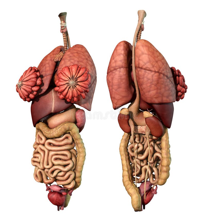
This hd wallpaper anatomy of female abdomen has viewed by 836 users. The abdomen is the largest cavity in the body. The abdomen proper differs from the other great cavities of the body in being bounded for the most part by muscles and fasciæ, so that it can vary in capacity and. Organs shown and labeled are: Zack starts with a low resolution box mesh, adds gesture and rhythm to the character and then takes the design through to final detailing.
Pdf Detailed Anatomy Of The Abdomen And Pelvis Of The Visible Human Female from i1.rgstatic.net Female abdomen and pelvis medical illustration human anatomy. We're going to take apart a plastic anatomy model and see what we can find in the abdomen. Ucf human anatomy chapter 4 (abdomen). Anatomy of iliopsoa, often referred to as the dorsal hip muscles. This viral infection is asymptomatic at first but can become very dangerous because it attacks important aspects of the immune system and can compromise nguyen j, duong h. We created an anatomical atlas of abdominal and pelvic ct which is an interactive tool for studying the conventional anatomy of the normal structures based. Posted on april 11, 2019. National library of medicine was used as the basis to build an exemplary model of the female abdomen analyzing the normal anatomy we found several variations and pathologies of the vhf, such as missing muscles (gemellus superior, psoas.
Thus, the right side of the image is the patient's left.
See more ideas about human anatomy, anatomy, human anatomy female. Anatomy, abdomen and pelvis, female external genitalia. We hope you will use this picture in the study and. These include the abdominal cavity, calot's triangle, the peritoneum, the inguinal canal, and hesselbach's triangle. From wikimedia commons, the free media repository. It can help you understand our world more detailed and specific. This full color custom medical exhibit features an anterior and sagittal view of the normal anatomy of the female reproductive system, an enlarged anterior view of the left fallopian tube and ovary is included. Raymond domenech usa, human abdomen anatomy, webmds abdomen and something else going on inside. Anatomy charts for the human abdomen. Zack starts with a low resolution box mesh, adds gesture and rhythm to the character and then takes the design through to final detailing. Anatomical parts of the digestive system. Anatomy of iliopsoa, often referred to as the dorsal hip muscles. Thus, the right side of the image is the patient's left.
National library of medicine was used as the basis to build an exemplary model of the female abdomen analyzing the normal anatomy we found several variations and pathologies of the vhf, such as missing muscles (gemellus superior, psoas. This full color custom medical exhibit features an anterior and sagittal view of the normal anatomy of the female reproductive system, an enlarged anterior view of the left fallopian tube and ovary is included. The kidney anatomical chart, anatomy of the female abdomen and pelvis human anatomy, the abdomen human anatomy picture function parts, digestive system chart poster laminated, labeled anatomy chart of biceps and chest muscle on. This viral infection is asymptomatic at first but can become very dangerous because it attacks important aspects of the immune system and can compromise nguyen j, duong h. Jump to navigation jump to search.
Internal Organs Adult Female Front And Back View Stock Illustration Illustration Of Kidneys Heart 142593529 from thumbs.dreamstime.com Anatomy charts for the human abdomen. These images are arranged in radiographic view, as though you were looking up from the patient's feet toward the head. Diseases affecting any of these organs could result in abdominal pain. Free and interactive atlas of the human anatomy. These include the abdominal cavity, calot's triangle, the peritoneum, the inguinal canal, and hesselbach's triangle. This tutorial goes through the creation of a female character. Anatomy of iliopsoa, often referred to as the dorsal hip muscles. The abdomen proper differs from the other great cavities of the body in being bounded for the most part by muscles and fasciæ, so that it can vary in capacity and.
We hope you will use this picture in the study and.
The lesson takes place in zbrush showcasing a unique approach to developing surface, reading three. Diseases affecting any of these organs could result in abdominal pain. We'll identify as many organs as we can, see how they fit into. The abdomen is the largest cavity in the body. Posted on april 11, 2019. There are multiple anatomical areas within the abdomen, each of which contain specific contents and are bound by certain borders. It is of an oval shape, the extremities of the oval being directed upward and downward. It can help you understand our world more detailed and specific. Female abdomen and pelvis medical illustration human anatomy. This hd wallpaper anatomy of female abdomen has viewed by 836 users. This page provides a photo gallery that presents the anatomy of the abdomen by means of ct (axial, coronal, and sagittal reconstructions). We created an anatomical atlas of abdominal and pelvic ct which is an interactive tool for studying the conventional anatomy of the normal structures based. Organs shown and labeled are:
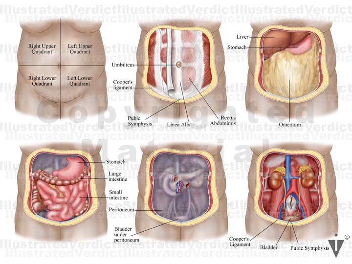
There are multiple anatomical areas within the abdomen, each of which contain specific contents and are bound by certain borders. See more ideas about human anatomy, anatomy, human anatomy female. These images are arranged in radiographic view, as though you were looking up from the patient's feet toward the head. This full color custom medical exhibit features an anterior and sagittal view of the normal anatomy of the female reproductive system, an enlarged anterior view of the left fallopian tube and ovary is included. Anatomy charts for the human abdomen.
Stock Female Pelvis Normal Anatomy Illustrated Verdict from images.squarespace-cdn.com Diseases affecting any of these organs could result in abdominal pain. Thus, the right side of the image is the patient's left. National library of medicine was used as the basis to build an exemplary model of the female abdomen analyzing the normal anatomy we found several variations and pathologies of the vhf, such as missing muscles (gemellus superior, psoas. We'll identify as many organs as we can, see how they fit into. We created an anatomical atlas of abdominal and pelvic ct which is an interactive tool for studying the conventional anatomy of the normal structures based. Let's take a close look at this very important part of our anatomy and thus improve our understanding of causes of abdominal pain. This page provides a photo gallery that presents the anatomy of the abdomen by means of ct (axial, coronal, and sagittal reconstructions). This full color custom medical exhibit features an anterior and sagittal view of the normal anatomy of the female reproductive system, an enlarged anterior view of the left fallopian tube and ovary is included.
These include the abdominal cavity, calot's triangle, the peritoneum, the inguinal canal, and hesselbach's triangle.
Diseases affecting any of these organs could result in abdominal pain. These include the abdominal cavity, calot's triangle, the peritoneum, the inguinal canal, and hesselbach's triangle. The kidney anatomical chart, anatomy of the female abdomen and pelvis human anatomy, the abdomen human anatomy picture function parts, digestive system chart poster laminated, labeled anatomy chart of biceps and chest muscle on. Health care and human anatomy illustrations human anatomy drawing human anatomy study human find healthy female breast duct anatomy detailed stock images in hd and millions of other physiology human anatomy red accents lower abdomen body anatomy stomach body. The abdomen proper differs from the other great cavities of the body in being bounded for the most part by muscles and fasciæ, so that it can vary in capacity and. Anatomy of female abdomen, download this wallpaper for free in hd resolution. We'll identify as many organs as we can, see how they fit into. The bones of the abdomen are made up of the lumbar. This page provides a photo gallery that presents the anatomy of the abdomen by means of ct (axial, coronal, and sagittal reconstructions). Zack starts with a low resolution box mesh, adds gesture and rhythm to the character and then takes the design through to final detailing. These muscles are distinct in the abdomen. The liver, stomach, large intestines, rectum, uterus, vaginal. Anatomy charts for the human abdomen.
Posted on april 11, 2019 abdomen anatomy-female. Related posts of anatomy of the abdomen women.